The shoulder is one of the most common areas of complaint that I see at the office. After necks and low backs, it's probably tied with feet for the body part that causes the most trouble. As an aside...yes, chiropractors work on more than just necks and backs! If a muscle or a joint is involved, chances are I've had a patient with the issue you may have. Today I'd like to look at some of the anatomy of the shoulder.
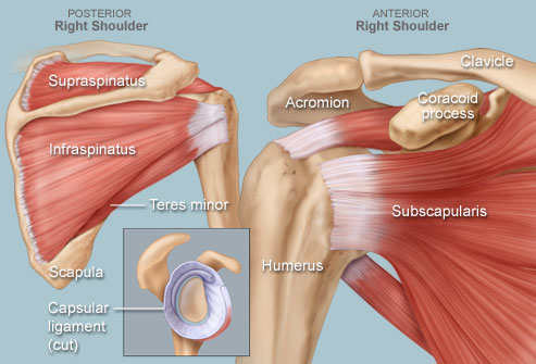
The bones that form the pectoral (shoulder) girdle on each side of the body are the scapula (also known as the shoulder blade) and clavicle (collar bone) whereas the glenohumeral (shoulder) joint is formed by the intersection of the head of the humerus and glenoid cavity of the scapula. This coming together of the humerus and scapula is assisted by a narrow rim of cartilage known as the glenoid labrum, which slightly enlarges the glenoid cavity in order to better hold the humeral head in place. The clavicle and scapula form joints at the top of the shoulder at the acromioclavicular joint (so named because the acromion of the scapula meets the acromial extremity of the clavicle).
The shoulder joint is a ball-and-socket joint that allows the upper arm to have a whole range of movement including flexion (bending up), extension (straightening out), abduction (movement away from the body), adduction (movement towards the body), lateral rotation (rotation of the arm at the shoulder joint itself) and circumduction (making circles with the arm). The shoulder joint allows a greater range of movement than any other joint in the body, and is encapsulated by a loose sac called the articular capsule that covers the area from the glenoid cavity to the neck of the upper humerus. Three sets of ligaments hold the scapula and humerus together. These are made up of the coracohumeral ligament, glenohumeral ligaments and transverse humeral ligament. Although the ligaments lend some degree of support to the shoulder joint, the majority of its strength actually comes from the associated muscles and tendons. Muscles of the shoulder and their tendons encircle the shoulder joint and form what is known as the rotator cuff. In practice, I've heard this called many things, from "Roller Cup" to "Rotator Cusp". The rotator cuff is actually four individual muscles working together to join the articular capsule and, together with the shoulder ligaments, hold the scapula and humerus in place. Four strategically located bursae (the subscapular, subdeltoid, subacromial and subcoracoid) prevent friction between the shoulder bones as they are moved by muscular contractions.
Although part of the axial skeleton of the torso, the shoulder girdle is not physically connected to the vertebral column but held in place by a series of muscles that include the trapezius, deltoid, serratus, rhomboid, subclavius, pectoralis and latissimus dorsi.
Phew...that was a lot of big words! Every single one of those parts of shoulder anatomy play an important role in the health of the shoulder. Often, surgery will look to remove or 'fix' one specific muscle or ligament, and often it is helpful. Looking at the entirety of the shoulder, and the surrounding parts of the body (like the neck and midback) is just as important, though. More often than not, where the pain is showing up is not where the actual problem is occurring. A famous medical doctor from Eastern Europe, Dr. Karl Lewitt, once said, "If you are focusing on where the pain is, you are lost."
Keep that in mind when it comes to your shoulder pain. While a very complicated structure with many parts that can 'fail', it's often just the weakest link in a chain of events that reach far beyond the shoulder.

No comments:
Post a Comment